-

- Exhibitors 2023
-
List of exhibitors
Advertisement
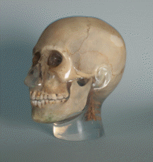
The X-ray head phantom was developed to simulate X-ray scenarios under real conditions in a training or research environment.
The complete skull structure of the phantom is embedded in tissue-equivalent material. The jaw is slightly open, so that the course of the teeth can be imaged realistically (for example, in panoramic x-ray exposures).
The neck section features five cervical vertebrae. An embedded thread at the bottom enables the phantom to be mounted on a standard tripod.
Hall 10.1 | F070